In-house diagnostic imaging allows quick and accurate diagnoses of your pet condition. MVP offer
- CT scanning
- Ultrasonography
- Echocardiogram
- Digital Radiography
CT (Computed Tomography)
CT is a highly advanced form of imaging that uses X-rays to create detailed cross-sectional images of the body. It allows us to see structures in far greater detail than standard X-rays.
CT is particularly useful for:
- Oncological/ cancer diagnosis and treatment planning
- Orthopaedic issues: joint disease (i.e. elbow dysplasia) and complex fractures
- Soft tissue and foreign bodies: detecting internal bleeding, organ abnormalities, or migrating foreign bodies
- Neurological conditions: examining the brain and spinal cord for disease (i.e. slipped discs)
- Head issues: dental disease, skull fractures, nasal disease (i.e. rhinitis/tumours) and jaw problems
- Respiratory and Thoracic studies: diagnosing chronic coughing, breathing difficulties and lung disease
Because CT provides 3D imaging, it helps us diagnose conditions more accurately and plan treatment with greater precision. Most pets require a general anaesthetic or sedation to keep them completely still during the scan, ensuring the best quality images. Some pets will have an intravenous contrast study to allow improve organ or tumour assessment.
Digital radiography
Radiography is an essential diagnostic tool used every day in practice. It allows us to quickly assess bones, joints, the chest, abdomen, and more.
We commonly use X-rays to:
- Diagnose fractures (broken bones) and joint problems
- Investigate lameness
- Assess heart and lung conditions
- Looking for bladder stones
- Checking abdominal organs
Our digital system produces high-quality images within seconds, allowing our vets to assess them immediately. This speeds up diagnosis and means we can discuss findings with you without delay.
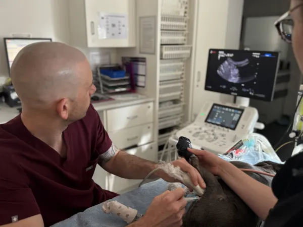
Ultrasonography
Ultrasonography uses sound waves, not radiation to create real-time images of the internal organs. It is particularly useful for examining soft tissues that may not be visible on X-rays.
Ultrasound helps us assess:
- Liver, kidneys, and bladder
- Spleen and pancreas
- Pregnancy scans
- Abdominal masses
- Fluid within the abdomen or chest
Echocardiogram is a non-invasive ultrasound scan that use sound waves to produce, real-time, moving images of the heart structure, chambers, values and blood flow. Usually completed in under 60minutes with your pet awake. This test allows for assessment, diagnosis and treatment planning, along with long-term treatment monitoring to assess heart function, damage and disease.
Why Advanced Imaging Matters
Having CT, digital radiography, and ultrasound available within our practice allows us to:
- Diagnose complex conditions sooner
- Reduce referral delays where appropriate
- Accurate surgical planning
- Improve patient safety
- Provide a higher level of care locally
Our experienced veterinary team carefully selects the most appropriate imaging modality for each individual case and will always discuss findings and treatment options with you clearly and compassionately.
In addition to supporting our own patients, we are pleased to offer outpatient referral imaging services for other veterinary practices. This means your vet can refer your pet to us specifically for CT, radiography, or ultrasound investigations, with results and reports shared promptly so ongoing care can continue seamlessly with your primary practice.
If your pet requires further investigation, whether as part of their care with us or via referral, our team will guide you through every step of the process. Please do not hesitate to Contact us

